hypokinetic lv | hypokinesis of left ventricle cause hypokinetic lv Left ventricular dysfunction is the medical name for a weak heart pump. It's a condition that impacts about 9% of people over the age of 60, which is around 7 million Americans. In this Mayo Clinic Minute, Dr. Paul Friedman, a Mayo Clinic cardiologist, explains what the condition is and how it can be diagnosed and treated. Datejust 36. Oyster, 36 mm, Oystersteel, Everose gold and diamonds. Reference 126281RBR. View variations. Make a date of a day. This Oyster Perpetual Datejust 36 in Oystersteel and Everose gold features a slate, diamond-set dial and a Jubilee bracelet. Slate Dial. A watchmaking technique.
0 · what is mild septal hypokinesis
1 · what does severely hypokinetic mean
2 · severe hypokinesis of the apex
3 · severe hypokinesis of left ventricle
4 · mild apical and septal hypokinesis
5 · hypokinesis of left ventricle cause
6 · hypokinesis of heart wall treatment
7 · causes of septal hypokinesis
The Rolex Datejust 41 has a diameter of 41 mm, and is thus the largest model in the series. It combines the popular elements found in the Datejust collection tracing back 70 .
Left ventricular dysfunction is the medical name for a weak heart pump. It's a condition that impacts about 9% of people over the age of 60, which is around 7 million .

st tropez gucci
Cardiac MRI showed a moderately dilated LV with mild-to-moderate concentric LV hypertrophy with wall thickness of 13 mm. LV systolic function was severely reduced with quantitative EF of . Mild hypokinesia basically means that the muscle of your heart does not contract as much as most peoples' hearts do. This may sound scary, but, do not be too worried because your ejection. Left ventricular dysfunction is the medical name for a weak heart pump. It's a condition that impacts about 9% of people over the age of 60, which is around 7 million Americans. In this Mayo Clinic Minute, Dr. Paul Friedman, a Mayo Clinic cardiologist, explains what the condition is and how it can be diagnosed and treated.

what is mild septal hypokinesis
We can see some hypokinesis of the anterior wall and overall mildly reduced heart function and ejection fraction. A reduced heart function and ejection fraction (EF) (<40%) usually manifests as fatigue and shortness of breath, sometimes even at rest.Cardiac MRI showed a moderately dilated LV with mild-to-moderate concentric LV hypertrophy with wall thickness of 13 mm. LV systolic function was severely reduced with quantitative EF of 22% and severe global hypokinesis.
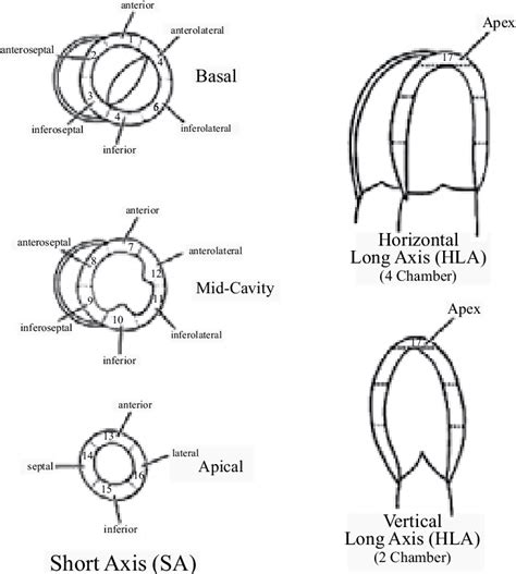
After anterior wall MI, reduced LVEF is a significant risk factor for thrombus formation, and most thrombi occur in the area of apical wall motion abnormalities in hypokinetic, akinetic, or dyskinetic (aneurysm) segments. The term "takotsubo" is taken from the Japanese name for an octopus trap, which has a shape that is similar to the systolic apical ballooning appearance of the LV in the most common and typical form of this disorder (movie 1 and movie 2); mid and apical segments of the LV are hypokinetic/akinetic, and there is hyperkinesis of the basal walls. The prognosis of left ventricular noncompaction (LVNC) remains elusive despite its recognition as a clinical entity for >30 years. We sought to identify clinical and imaging characteristics and risk factors for mortality in patients with LVNC.Standardized myocardial segmentation and nomenclature for echocardiography. The left ventricle is divided into 17 segments for 2D echocardiography. One can identify these segments in multiple views. The basal part is divided into six segments of 60° each.
what does severely hypokinetic mean
severe hypokinesis of the apex
The most common wall motion abnormality in NSC due to SAH appears to be either hypokinesis of the basal and midventricular segments with sparing of the apex or global LV hypokinesis, whereas the TC pattern is less common. 54–58,62 In a recent report of consecutive patients with NSC, 8 of 24 (33%) had isolated apical and midventricular LV .

When LV systolic function is impaired, the report will indicate whether the chamber was globally hypokinetic, typical of cardiomyopathy, or whether regional wall-motion abnormalities were seen, the result of myocardial infarction.
Mild hypokinesia basically means that the muscle of your heart does not contract as much as most peoples' hearts do. This may sound scary, but, do not be too worried because your ejection.
Left ventricular dysfunction is the medical name for a weak heart pump. It's a condition that impacts about 9% of people over the age of 60, which is around 7 million Americans. In this Mayo Clinic Minute, Dr. Paul Friedman, a Mayo Clinic cardiologist, explains what the condition is and how it can be diagnosed and treated. We can see some hypokinesis of the anterior wall and overall mildly reduced heart function and ejection fraction. A reduced heart function and ejection fraction (EF) (<40%) usually manifests as fatigue and shortness of breath, sometimes even at rest.Cardiac MRI showed a moderately dilated LV with mild-to-moderate concentric LV hypertrophy with wall thickness of 13 mm. LV systolic function was severely reduced with quantitative EF of 22% and severe global hypokinesis. After anterior wall MI, reduced LVEF is a significant risk factor for thrombus formation, and most thrombi occur in the area of apical wall motion abnormalities in hypokinetic, akinetic, or dyskinetic (aneurysm) segments.
The term "takotsubo" is taken from the Japanese name for an octopus trap, which has a shape that is similar to the systolic apical ballooning appearance of the LV in the most common and typical form of this disorder (movie 1 and movie 2); mid and apical segments of the LV are hypokinetic/akinetic, and there is hyperkinesis of the basal walls. The prognosis of left ventricular noncompaction (LVNC) remains elusive despite its recognition as a clinical entity for >30 years. We sought to identify clinical and imaging characteristics and risk factors for mortality in patients with LVNC.
Standardized myocardial segmentation and nomenclature for echocardiography. The left ventricle is divided into 17 segments for 2D echocardiography. One can identify these segments in multiple views. The basal part is divided into six segments of 60° each. The most common wall motion abnormality in NSC due to SAH appears to be either hypokinesis of the basal and midventricular segments with sparing of the apex or global LV hypokinesis, whereas the TC pattern is less common. 54–58,62 In a recent report of consecutive patients with NSC, 8 of 24 (33%) had isolated apical and midventricular LV .
severe hypokinesis of left ventricle
mild apical and septal hypokinesis
$12K+
hypokinetic lv|hypokinesis of left ventricle cause


